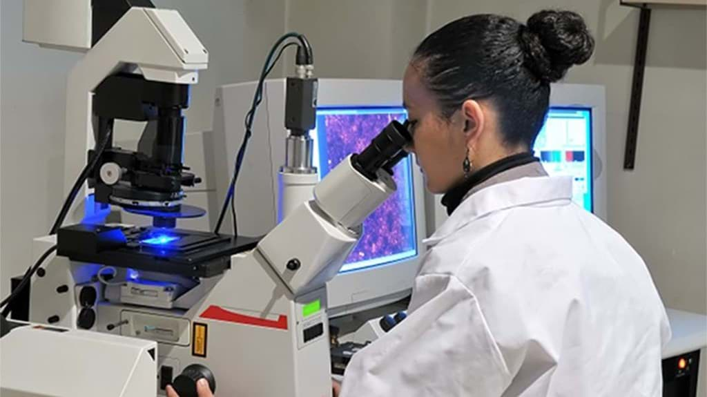
5 ways you don't have to rely on a fluorescent microscopy expert ever again
How a multicolor fluorescence microscope will make you go bonkers.
Remember the moment you worked a whole week preparing your cells to end up behind a microscope you do not know how to use? Frantically looking for someone with expertise, but to find no one available?
Learn more about
In summary?
- What is the problem with conventional microscopy?
- How does it affect your work?
- How can you prevent this and be a microscopy expert?
- Our solutions to become an expert.
What is the problem?
What is the problem with conventional microscopy?
You are sitting uncomfortably in a dark room because the system is too complicated and destined to ruin the images you need to publish your work.Conventional microscopes have a multitude of functionalities requiring expertise to get your experiments going. In most institutes and companies, a multicolor microscope is found somewhere at a foreign lab in a dark backroom with personnel you don't know. Honestly, none of them are interested in helping you.
The rapid advancement of new microscopy techniques tremendously improved the visualization of biological processes. However, these new techniques challenge the use of the system. They often come with an overwhelming set of parameters and settings, which are hard to understand and, in addition, to reproduce.

Effect on research
How does it affect your work?
Getting the optimal settings for your fluorescence microscope publishable image is often time-consuming and challenging, especially for inexperienced users. To reproduce the acquired image in your following experiment or that of another researcher, the parameters and settings should be well-defined and logged, if not better, saved on the device. Due to its complex nature, writing down the settings is often neglected. Or not clearly mentioned in the material and methods.We often end up with unusable data that is not reproducible. Or worst, it is not admissible in a paper.
It does not sound gratifying when it should be the climax of your work to look at the biological processes you are so keen to unravel. Surely, we don't want to leave this extraordinary moment in the hands of a microscope operator.
Expert in microscopy
How can you prevent this and be a microscopy expert?
Technology has advanced, making microscopy complicated. On the bright side, innovation and more manageable choices are also at our disposal. These tips help you be a professional yourself.- Stop wasting time translating protocols to difficult settings and not getting it right. An all-in-one fluorescence microscope with an onboard computer and straightforward analysis software is all you need. With just a few mouse clicks, you can generate time-lapse and Z-stack images in a flash.
- Ban human errors with straightforward analysis software. Work consistently and get reproducible data. Leave the complicated matters to the machine. For instance, let the software automatically merge images.
- Especially cell culture labs benefit from all-in-one devices, including easy-to-set-up life cell imaging, time laps, image editing, and cell counting protocols. And easy to adjust settings. Even students should be able to work effortlessly.
- Select a multicolor microscope with no difficult or indistinct parameters. Save time with software enabling you to choose from many measurement and annotation tools. Go back and forth or modify your protocol with ease.
- Get rid of complex system setups resulting in an uncomfortable and incomprehensible workspace. You can have complicated workspaces on your bench, like life cell imaging, and access them on a single instrument every minute of the day.
Our solutions
Our solution to become an expert
You can have all the perks of a conventional multicolor fluorescence microscope, like a scientific-grade CMOS camera and five proven quality objective lenses of choice, without requiring technical knowledge to use high-end features to capture high-quality images. The CELENA S digital imaging system is user-friendly, requiring no training to set up imaging protocols. Even students learn their way around in minutes with this all-in-one device.
Allow yourself to capture vivid, publishable images easily. You can use the CELENA S for multiple applications, such as capturing and analyzing multicolor fluorescence images, live cell imaging, and automated cell counting. With the onboard data analysis, study your images immediately upon capture. Never lose your protocols and measurement data by saving it readily to a USB drive.
The addition of the fibration isolation chamber eliminates complex microscopical setups. Control the temperature, humidity, and gas content precisely at your lab bench.
Z-stack imaging is not only for professionals any longer. With this digital imaging system, you can master this seemingly complex cell capture technique.
For high-end users, the CELENA X fully automated high-content imager offers all the perks of the CELENA S. It automatically captures crisp images for various applications, including live-cell imaging, and is optimized for fully automated plate and slide imaging.
You can capture images in multicolor fluorescence, brightfield, color brightfield, and phase contrast and customize the CELENA X to your needs.
Lorem
Success stories
Lorem ipsum dolor sit amet, consectetur adipiscing elit. Nunc nec nibh sed sem mattis malesuada. Aenean lobortis vulputate massa sit amet pellentesque. Duis quam sapien, fermentum nec porta vitae, laoreet sed nunc. Suspendisse scelerisque arcu eu molestie mattis. Ut ornare sit amet ante viverra luctus. Integer eleifend sollicitudin purus, eget suscipit massa blandit in. Fusce neque tortor, laoreet vel justo vitae, mollis dignissim ex. Morbi quis ultrices nulla, eget accumsan augue. Sed ut lacus vitae velit tincidunt mollis ac eu sapien. Phasellus ullamcorper est quis massa pharetra, sit amet molestie mi molestie. Etiam laoreet, leo ac tincidunt commodo, elit eros eleifend nisi, sit amet convallis nulla libero id lacus. Vivamus facilisis gravida varius. Praesent efficitur venenatis augue et efficitur.
Our news
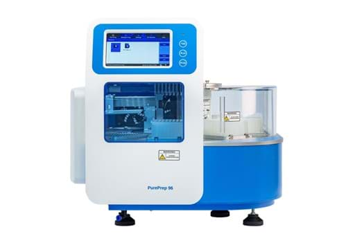
Innuscreen introduces PurePrep benchtop systems for autom...
Discover the PurePrep family for quick and effective DNA/RNA extraction.
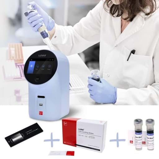
Is more sustainable cell counting possible?
This article discover eco-friendly solutions for cell counting
Introducing Phoenix Instrument: small lab equipment now a...
January 2025 - Phoenix Instrument | Small lab equipment now available at Westburg Life Sciences


